Spleen
The spleen is being the largest single mass of lymphoid tissues in the body.
Spleen is located in the left hypochondrium opposite the levels of the 9th and 11th ribs; its long axis is parallel to the 10th rib.
It is a wedge-shaped organ. It is about 1 inch thick, 3 inches long,7 ounces in weight, and is related to the 9tha to 11th ribs.
Structure of spleen
1. Outer serous layer, made up of peritoneum
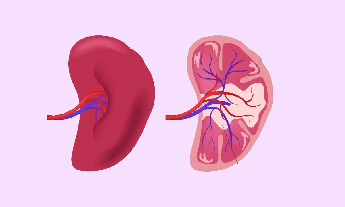
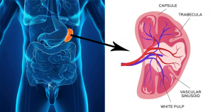
2. Deep to it, a thick fibrous capsule. The capsule sends numerous trabeculae into the substance of the spleen.
3. The white pulp: It is made of lymphatic tissue, which forms dense collections around the arterioles. They form cord-like aggregations and follow the branching pattern of the arterioles.
4. The red pulp: It fills the rest of the spleen contains blood, RBC, macrophages, and lymphocytes.

External features of spleen
The spleen has two ends,3 borders, and 2 surfaces.
The superior border shows notches: one of them, called the splenic notch, is prominent; it lies near the anterior end.
The outer, diaphragmatic surface of the spleen is convex and smooth. The visceral surface shows the impressions of adjacent viscera [a]the gastric impression produced by the fundus of the stomach, which lies in between the superior and intermediate borders:[b]the renal impression, for the left kidney, is seen between the intermediate and inferior borders,[c]Colic impression for the splenic flexure of the colon; close to the anterior end of the spleen;[d]the pancreatic impression; for the tail of pancreas, close to the hilum of the spleen.
The hilum of the spleen is inferomedial to the gastric impression. It transmits the splenic vessels and nerves. It also provides attachment to the gastrosplenic and lienorenal ligaments.
The diagram separates the diaphragmatic surface of the spleen from the left costodiaphragmatic recess, the lung, and ribs.
Blood Supply
Arterial supply
The arterial supply of the spleen is by the splenic artery, the largest branch of the coeliac trunk. It is tortuous; it passes through the lienorenal ligament to the hilum, where the artery divides into 4-5 branches.
Drainage of the veins
The splenic vein arises from the spleen’s hilum. The portal vein is formed when it joins the superior mesenteric vein behind the pancreas’ neck.
Functions of Spleen
- Both B-Lymphocytes and T-lymphocytes multiply in the spleen and play an important role in immune responses.
- Spleen is considered to be an important component of the reticuloendothelial system. It contains a large number of macrophages. Their main function is the destruction of RBCs that have completed their life span. The macrophages also destroy worn-out leukocytes and bacteria.
- Hemopoiesis, during fetal life.

![Cumin[Jeera]Water For Weight Loss: Facts Behind It !](https://medicalfitbit.com/wp-content/uploads/2021/10/Water-For-Weight-Loss.jpg)
![Breast Cancer and symptoms: [Anatomical Features]](https://medicalfitbit.com/wp-content/uploads/2021/10/Breast-Cancer.jpg)