Heart
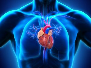
The heart is placed in the middle mediastinum in the thorax. It lies between the two lungs and immediately above the diaphragm. It is situated behind the sternum and adjoining costal cartilages and left ribs.
average weight: Males -300gm;Female-250gm
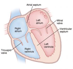
Chambers of Heart
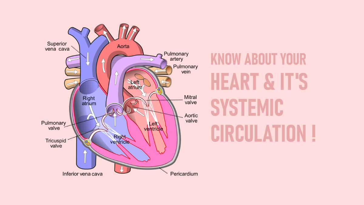
The heart has 4 chambers-2atria[the receiving area]and 2 ventricles, the discharging chambers.
The wall of each chamber consists of 3 layers:
1. An internal layer or endocardium
2. A middle layer or myocardium composed of cardiac muscles.
3. An outer layer or epicardium
The myocardium is the thickest layer and forms the main mass of the heart
Base, Apex and surfaces
The heart has a base, an apex and 3 surfaces-sternocostal ,the diaphragmatic and pulmonary surfaces :it has 4 boarders-right ,left, superior and inferior borders.
The base of the heart
The base is situated posteriorly and is formed mainly by the left atrium [the heart does not rest on its base].
The apex of the heart
The blunt apex is formed by the left ventricle . It is located posterior to the 5th left intercostal space in adults, just medial to the midclavicular line[7-9cm from the median plane].
The diaphragmatic [inferior]surface of the heart is formed by both ventricles, mainly the left one. It is related to the central tendon of the diaphragm. The posterior interventricular groove divides this surface into right one-third and left two-thirds.
Borders
The right border is made by the right atrium, the inferior border is formed mainly by right ventricle and partly the left; the left border is formed is formed mainly by left ventricle and partly by left auricle. The great vessels enter and leave the superior border of the heart.
External and internal features of chambers
Right atrium
It is the right upper chamber of the heart.Right atrium forms the right border, part of the upper border, the sternocostal surface, and part of the base of the heart . It receives blood from the entire body and pumps it to the right ventricle through the right atrioventricular or tricuspid orifice[opening].
External features
It receives the superior vena cava at the upper end and the inferior vena cava at the lower end. The upper end shows a projection to the left side, the auricle. Its margins are notches, and the interior is rough and sponge-like. The auricle partly covers and partly overlaps the infundibulum of the right ventricle and partly covers and partly overlaps the root of the ascending aorta.
There is Extending from the superior vena cava to the inferior vena cava, along the right border of the atrium, is a shallow vertical groove-the sulcus terminalis.
The atrioventricular groove or coronary sulcus separates the right atrium from the right ventricle. This groove houses the right coronary artery and the tiny cardiac vein.
Veins draining into the right atrium
1. Superior vena cava
2. Inferior vena cava
3. Coronary sinus
4.Anterior cardiac veins
5. Venae cordis minimi
Internal features
The right atrium has three parts:
1. sinus venarum
2. The rough anterior part or pectinate part, including the auricle
3. The interatrial septum
Right Ventricle
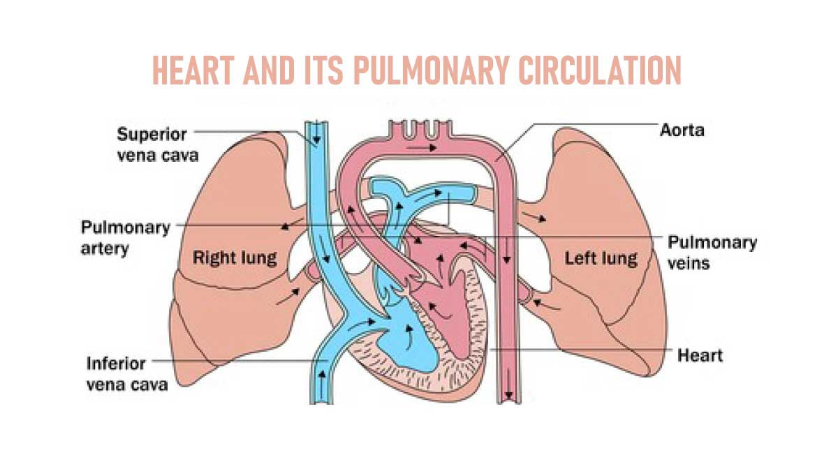
The right ventricle forms the inferior border and a large part of the sternocostal surface of the heart. It is a triangular chamber. The pulmonary trunk and pulmonary arteries take deoxygenated blood from the right atrium and pump it to the lungs.
Interior of the right ventricle
It has an inflowing part and an outflowing part.
Inflowing part
It is rough due to the presence of muscular ridges called trabaculae carneae.
Outflowing part
The outflowing part or infundibulum is smooth. It forms the upper conical part of the right ventricle which gives origin to the pulmonary trunk.
The interior of the right ventricle shows two orifices:
1. The right atrioventricular orifice or tricuspid orifice, guarded by the tricuspid valve.
2. the pulmonary orifice is guarded by the pulmonary valve.
The interior of the inflowing part shows trabaculae carneae or muscular ridges of 3 types.
1. Ridges or fixed elevations
2. Bridges
3. Papillary muscles or pillars with cordae tendinae connecting one end to the ventricular wall and the other end to the tricuspid valve cusps.
Left Atrium
It represents the left two-thirds of the base of the heart, the greater part of the upper border, parts of the sternocostal and left surfaces, and part of the left border of the heart.
It is a quadrangular chamber receiving oxygenated blood from the lungs through four pulmonary veins and pumps to the left ventricle through the left atrioventricular or bicuspid or mitral orifice, which is guarded by the mitral valve[bicuspid valve].
The appendage of the left atrium, the left auricle, projects anteriorly to overlap the infundibulum of the right ventricle.
On each side of the posterior wall, two pulmonary veins enter into the left atrium. Musculi pectinate is present only in the auricle. The anterior wall of the atrium is formed by the interatrial septum. A few venae cordis minimi open directly into the left atrium.
Left Ventricle
The left ventricle gathers and pumps oxygenated blood from the left atrial into the aorta.
It forms the apex of the heart, a part of the sternocostal surface, most of the left border and left surface, and the left two-thirds of the diaphragmatic surface.
The interior of the left ventricle has two parts:
1. The lower rough part with trabaculae carneae
2. The smooth upper part or aortic vestibule gives origin to the ascending aorta.
The interior of the ventricle shows two openings:
1. The left atrioventricular or bicuspid, or mitral orifice is guarded by the mitral valve.
2. The aortic orifice, guarded by the aortic valve.
Blood Supply
Arterial supply
The heart gets its oxygen and nutrients from 2 arteries-the right and left coronary arteries, which are the first branches of aorta.
The right coronary artery[RCA]
It occurs from the right aortic sinus and descends in the coronary sulcus between the right auricle and right ventricle. It then passes towards the inferior border of the heart, where it gives off a marginal branch that runs towards the apex. After giving off this branch, the RCA turns to the left and enters the posterior interventricular groove, where it gives off its largest branch, the posterior interventricular branch. This branch supplies both ventricles runs towards the apex and anastomoses with the anterior interventricular branch of the left coronary artery.
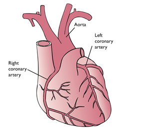
The left coronary artery[LCA]
It arises from the left aortic sinus; passes between the left auricle and pulmonary trunk to reach the coronary groove. It soon divides into two terminal branches[1], The anterior interventricular branch and[1]the circumflex branch.
The anterior interventricular branch passes along the anterior interventricular groove to the apex of the heart; It anastomoses with the right coronary artery’s posterior interventricular branch.
![Breast Cancer and symptoms: [Anatomical Features]](https://medicalfitbit.com/wp-content/uploads/2021/10/Breast-Cancer.jpg)